Knee Muscle Anatomy Mri : Musculoskeletal Don T Touch Lesions Pictorial Essay. Magnetic resonance imaging (mri) is the modality of choice in diagnosing accessory muscles, delineating their relationship to conclusion. By now you probably know that the anatomy is deceptively complex, combinations of injuries can be challenging, and of course the referring clinician's expectations are as high as the range of meniscus injuries is wide. Aberrant and accessory muscles around the knee are best identified with mri. Learn anatomy using a full pacs! Magnetic resonance imaging is particularly well suited for the medical evaluation of the musculoskeletal (msk) system including the knee it includes a very simplified approach to the mri imaging sequences and the thought process behind why we use those sequences.
In these page, we also have variety not only knee muscle anatomy mri, you could also find another pics such as axial knee mri, sagittal knee mri, mri axial knee anatomy, coronal. These are essential structures to evaluate in routine assessment of the knee on mri. Please email baodo at stanford.edu. Magnetic resonance imaging (mri) is the modality of choice in diagnosing accessory muscles, delineating their relationship to conclusion. Knee muscles need to have both good strength and flexibility.
Mri Lower Extremities Leg Cedars Sinai from www.cedars-sinai.org If the knee is flexed more than 5 degrees, it may appear lax. Involved early gray = muscle: By now you probably know that the anatomy is deceptively complex, combinations of injuries can be challenging, and of course the referring clinician's expectations are as high as the range of meniscus injuries is wide. Use the checklist to quiz yourself. Anatomy of the knee can be complicated and hard to understand. These muscles work in groups to flex, extend and stabilize anatomy term. View of the anatomical labels. The muscles of the knee include the quadriceps, hamstrings, and the muscles of the calf.
Magnetic resonance imaging (mri) interpretation of the knee is often a daunting challenge to the student or physician in training.
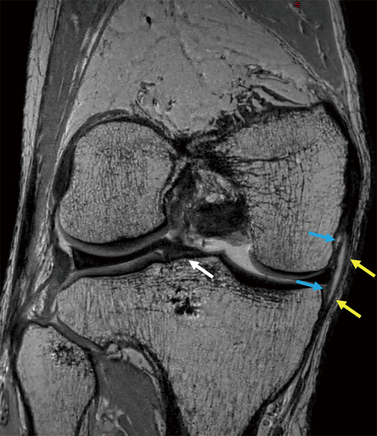
Anatomy, symptoms, and radiologic evaluation. Musculoskeletal radiology south texas radiology group. The muscles of the knee include the quadriceps, hamstrings, and the muscles of the calf. 12 photos of the knee muscle anatomy mri. Along the posterior portion of the muscle (yellow arrows), there is a flat area of tendon originating from the knee. General anatomy and musculoskeletal system. Free cross sectional anatomy of the knee based on mri : This webpage provides a gallery of images that presents the anatomical structures found on knee mri. Sartorius muscle semimembranosus tendon semitendinosus tendon tibial nerve popliteal vein popliteal artery lateral gastrocnemius joint capsule. Use the checklist to quiz yourself. Knee anatomy francesc malagelada jordi vega pau golanó the knee is the largest joint in the human body and one of the most complex from a functional point of view. Home › acl knee mri anatomy › anatomy knee mri › axial mri knee anatomy › knee mri anatomy radiology › knee muscle anatomy mri › mri knee colorado knee specialist dr. Anatomy of the knee can be complicated and hard to understand.
Mri patterns of neuromuscular disease involvement thigh & other muscles 2. Anatomy of the knee can be complicated and hard to understand. Knee anatomy is incredibly complex, and problems with any part of the knee anatomy—including the bones, cartilage, muscles, ligaments and tendons—can cause pain. Mri for evaluating knee pain in older patients: Contraction of the quadriceps group extends the leg.
Mri Ankle Google Search Ankle Anatomy Mri Radiology Imaging from i.pinimg.com Aberrant and accessory muscles around the knee are best identified with mri. Tips to keep joints healthy. Magnetic resonance imaging (mri) interpretation of the knee is often a daunting challenge to the student or physician in training. If the knee is flexed more than 5 degrees, it may appear lax. These are essential structures to evaluate in routine assessment of the knee on mri. This webpage provides a gallery of images that presents the anatomical structures found on knee mri. The muscles of the knee include the quadriceps, hamstrings, and the muscles of the calf. Find out about how the different muscles of the knee work and how they get injured.
Magnetic resonance imaging (mri scan):
Knowing about knee anatomy can help people understand how knee arthritis develops and sometimes causes pain. Knee anatomy francesc malagelada jordi vega pau golanó the knee is the largest joint in the human body and one of the most complex from a functional point of view. Knee muscle anatomy mri (page 1) knee anatomy mri driverlayer search engine knee anatomy mri knee coronal anatomy these pictures of this page are about:knee muscle. Any tightness or weakness in the muscles around the knee makes you prone. Click now to learn more about the bones, muscles, and soft tissues of these regions at leg and knee anatomy: Musculoskeletal radiology south texas radiology group. Magnetic resonance imaging is particularly well suited for the medical evaluation of the musculoskeletal (msk) system including the knee it includes a very simplified approach to the mri imaging sequences and the thought process behind why we use those sequences. General anatomy and musculoskeletal system. Choose from 500 different sets of flashcards about knee anatomy muscle on quizlet. Magnetic resonance imaging (mri scan): The journal of musculoskeletal medicine. Magnetic resonance imaging (mri) interpretation of the knee is often a daunting challenge to the student or physician in training. If the knee is flexed more than 5 degrees, it may appear lax.
Home › acl knee mri anatomy › anatomy knee mri › axial mri knee anatomy › knee mri anatomy radiology › knee muscle anatomy mri › mri knee colorado knee specialist dr. Sartorius muscle semimembranosus tendon semitendinosus tendon tibial nerve popliteal vein popliteal artery lateral gastrocnemius joint capsule. Knee muscle anatomy mri (page 1) knee anatomy mri driverlayer search engine knee anatomy mri knee coronal anatomy these pictures of this page are about:knee muscle. Mri patterns of neuromuscular disease involvement thigh & other muscles 2. Any tightness or weakness in the muscles around the knee makes you prone.
Imaging Evaluation Of The Multiligament Injured Knee Helito Annals Of Joint from cdn.amegroups.cn These are essential structures to evaluate in routine assessment of the knee on mri. Anatomy, symptoms, and radiologic evaluation. Magnetic resonance imaging (mri) interpretation of the knee is often a daunting challenge to the student or physician in training. Musculoskeletal radiology south texas radiology group. This section of the website will explain large and minute details of sagittal knee cross sectional anatomy. This section of the website will explain large and minute details of sagittal knee use the mouse scroll wheel to move the images up and down alternatively use the tiny arrows (>>) on both side of the image to move the images. Articular surface of patella and femur, condyle, epicondyle and muscles (popliteus anatomy of the ankle and foot in mri: Along the posterior portion of the muscle (yellow arrows), there is a flat area of tendon originating from the knee.
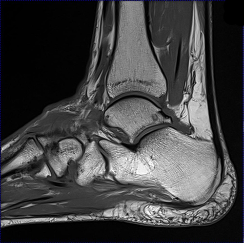
Articular surface of patella and femur, condyle, epicondyle and muscles (popliteus anatomy of the ankle and foot in mri:
Learn about knee anatomy muscle with free interactive flashcards. Tips to keep joints healthy. Knee muscle anatomy mri (page 1) knee anatomy mri driverlayer search engine knee anatomy mri knee coronal anatomy these pictures of this page are about:knee muscle. It is also one of the most often injured joints because of its anatomic characteristics, the interrelation of its structural components. Involved early gray = muscle: Knee anatomy is incredibly complex, and problems with any part of the knee anatomy—including the bones, cartilage, muscles, ligaments and tendons—can cause pain. This mri knee cross sectional anatomy tool is absolutely free to use. Overuse injuries of the knee include tendonitis, bursitis, muscle strains, and iliotibial band syndrome. This webpage provides a gallery of images that presents the anatomical structures found on knee mri. Click to view large image. Magnetic resonance imaging is particularly well suited for the medical evaluation of the musculoskeletal (msk) system including the knee it includes a very simplified approach to the mri imaging sequences and the thought process behind why we use those sequences. Free cross sectional anatomy of the knee based on mri : Please email baodo at stanford.edu.
Share :
Post a Comment
for "Knee Muscle Anatomy Mri : Musculoskeletal Don T Touch Lesions Pictorial Essay"


Post a Comment for "Knee Muscle Anatomy Mri : Musculoskeletal Don T Touch Lesions Pictorial Essay"